|
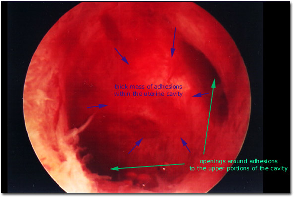
Photograph of the hysteroscopic view of a uterine cavity with severe intrauterine synechiae (Ashermans syndrome) that occupied the bulk of the cavity. The midline fundal location of the abnormal tissue is similar to that seen with a uterine septum (the eccentric rotation of the scar tissue is not characteristic of a uterine septum). These very thick intrauterine adhesions can be muscular and vascular, so the possibility of excessive absorption of hysteroscopic distending media during resection should be considered when performing hysteroscopic transection of these lesions.
|

|