|
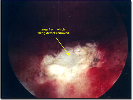
Photograph of the hysteroscopic view of a uterine cavity following removal of a broad based endometrial polyp. The edges of the (endometrial polyps) stalk that remain visible in this photograph would normally be removed with the loop electrode without difficulty. Bleeding can be encountered when these masses are removed and electrocautery can usually control hemostasis easily (note the small region of char created by cautery on the right side of this visual field at the edge of the remaining stalk)
|

|