|
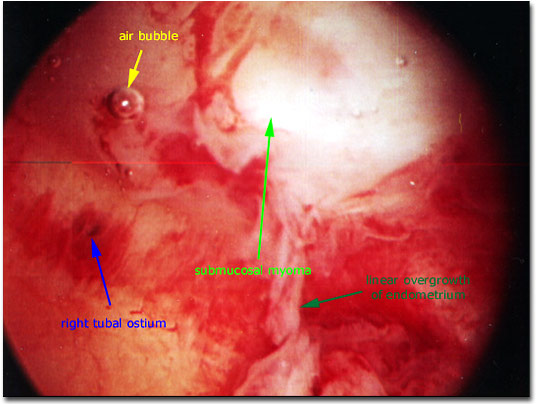
Photograph of a hysteroscopic view of the inside of a uterus that contains a 1-2 cm rounded submucosal myoma (white mass extending into the uterine cavity at the top of the visual field), a few small air bubbles in the liquid distending medium (here D5W), and the right tubal ostium (opening to the right fallopian tube) which is seen as a black dot off to the left side of the photograph midway between the top and the bottom of the picture.
|

|