|
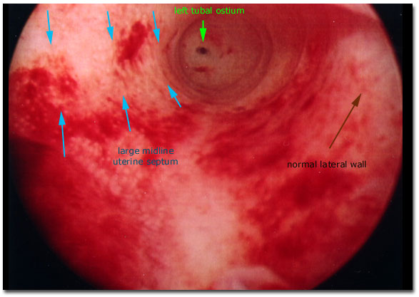
Photograph of the hysteroscopic view of a uterine cavity revealing the left tubal ostium (dark dot directly above the center of the photo), a normal appearing left lateral sidewall (of the cavity, to the right side of the visual field of view), and a mass of tissue occupying the space that normally exists between the right tubal ostium (not seen here) and the left tubal ostium. This tissue in the midline of the uterine cavity was determined to be a (relatively large) uterine septum.
|

|